Neurological Examination
Speech
Elements to test
1.
Comprehension
2.
Fluency
3.
Repetition
4.
Naming
5.
Quality
6.
Articulation/rhythm
Examination
- Ask patient
- Their name
- To describe
the room
·
Repeat “ No ifs ands or buts”
- Abnormal
with - Conductive or receptive
aphasia
- Repeat
- West-register
Street or British constitution
- Test
articulation/rhythm
- Repeat
- Puh – test
lips
- Tuh – test tongue
- Kuh – test
palate
- Staged
commands – one, two, three
- Tests comprehension (receptive) -
abnormal suggests receptive problem
·
Questions – further test comprehension – and hence receptive
- “Do you put
your socks on before your shoes?”
- “Do you
close the car door before you get in?”
- Word-finding/naming
- Name animals
in one minute NR18-22)
- All words
staring with a particular letter, usu f or s,
in 1 min NR>12)
- Name objects
getting more specific
- Watch –
dial, hands, Pen – tip,
- Abnormal
with all types of dysphasia however if other aspects normal except for
this then could be a specifically nominal
aphasia
- Jaw jerk
- Brisk if
pseudobulbar palsy
·
Further Ax
o
Look for facial asymmetry and potentially assess
facial nerve
o
Look for hemiparesis
o
Co-ordination or nystagmus – suggestive of
cerebellar dysfunction
Findings:
Dysphonia
·
Altered quality of the voice with normal fluency
but change in volume
- Vocal cord disease – e.g. recurrent
laryngeal nerve palsy
Dysarthria
·
Difficulty articulating
- DDX:
- LMN lesions
- Cranial
nerves – CNVII – slurred speech
- Bulbar
palsy – Nasal speech
- UMN lesions
- Pseudobulbar
palsy – spastic dysarthria – trying to squeeze out words
- Extrapyramidal
conditions
- Monotonous
speech
- Cerebellar
lesions
- Slow,
slurred, sometimes explosive speech
- May be
broken up into syllables – scanning speech
Aphasia/Dysphasia
- Wernike’s aphasia
- Poor
comprehension, fluent but meaningless.
No repetition.
- Broca’s aphasia
- Preserved
comprehension, non-fluent speech. No repetition.
- Conductive
aphasia
- Loss of
repetition with preserved comprehension and output.
- Transcortical
sensory aphasia
- As 1. but
with preserved repetition.
- Transcortical
motor aphasia
- As in 2. but
with preserved repetition
- Nominal apahsia
- Only
difficulty with naming
- Dominant
posterior temporoparietal lesion
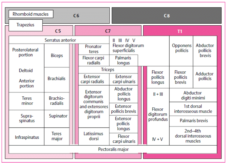
Upper Limb
|
Upper Limb |
|
|
|
|
|
Shoulder |
|
|
|
|
|
Abduction |
C5-C6 |
Axillary |
Deltoid |
|
|
Adduction |
C6-C7-C8 |
Thoracodorsal Pecoral nerves |
Latissimus
dorsi Pectoralis
major |
|
|
External
rotation |
C5-C6 |
Suprascapular |
Infraspinatus
|
|
|
Internal
rotation |
C5-C6-C7 |
Subscapular |
Subscapularis |
|
|
Elbow |
|
|
|
|
|
Flexion |
C5-C6
|
Musculocutaneous Radial |
Biceps
(when supine) Brachialis
(all positions) Brachioradialis
(in mid-position) |
C5-C6 C5-C6 C5-C6 |
|
Extension |
C6-C7-C8 |
Radial |
Triceps |
C6-C7-(C8) |
|
Wrist |
|
|
|
|
|
Flexion |
C6-C7-C8 |
Median Ulnar |
FCR Palmaris
longus FDS
(minor) FCU |
C6-C7 C7-C8 C7-C8 C7-C8 |
|
Extension |
C6-C7-C8
|
Radial |
ECR-
main muscle with normal wrist extension ECU |
C6-C7 C7-C8 |
|
Finger |
|
|
|
|
|
Flexion |
C7-C8 |
Median Ulnar |
FDP
– Digit 2-3, FDS Lumbricals FDP
– Digit 4, 5 Lumbricals |
C7-C8 C7-C8 C8-T1 C8-T1 C8-T1 |
|
Extension |
C7-C8 |
Radial |
Extensor
digitorum EIP |
C7 C7-C8 |
|
Abduction |
C8-T1 |
Ulnar |
Dorsal
interossei |
C8-T1 |
|
Adduction |
C8-T1 |
Ulnar |
Palmar
interossei |
C8-T1 |
|
Thumb |
|
|
|
|
|
Flexion |
C8-T1 |
Median |
FPL FPB |
C7-C8 C8-T1 |
|
Extension |
C8 |
Radial |
EPL |
C7-C8 |
|
Abduction |
C8-T1 |
Median |
APB |
C8-T1 |
|
Adduction |
C8-T1 |
Ulnar |
Adductor
pollicis |
C8-T1 |
Reflexes
|
Upper Limb |
|
|
Biceps |
C5-C6 |
|
Triceps |
C7-C8 |
|
Brachioradialis |
C5-C6 |
|
Finger |
|
Lower Limb
|
Lower Limb |
|
|
|
|
|
Hip |
|
|
|
|
|
Flexion |
L1-L2-L3-L4 |
Femoral
plexus Femoral
nerve |
Iliopsoas Rectus
femoris |
L2-L3 L2-L3 |
|
Extension |
L5-S1-S2 |
Inferior
gluteal |
Gluteus
maximus |
L5-S1 |
|
Abduction |
L4-L5-S1 |
Superior
gluteal nerve |
Gluteus
medius, Tensor fasciae latae |
|
|
Adduction |
L2-L3-L4 |
Obturator
|
|
|
|
Knee |
|
|
|
|
|
Flexion |
L5-S1-S2 (L5-S1) |
Sciatic |
Semimembranous/tendinous Biceps
femoris Adductors |
L4-L5-S1 L5-S1 L2-L4 |
|
Extension |
L2-L3-L4 |
Femoral |
Quadriceps |
L3-L4 |
|
Ankle |
|
|
|
|
|
Plantar
Flexion |
S1-S2 |
Tibial |
Gastrocnemius
Soleus |
L5-S1-S2 S1-S2 |
|
Dorsi
Flexion |
L4-L5 |
Deep
peroneal |
Tibialis
anterior |
L4-L5 |
|
Eversion |
L5-S1 |
Superficial
peroneal |
Peroneus
longus and brevis |
L5-S1 |
|
Inversion |
L5-S1 |
Tibial
|
Tibialis
posterior |
L5-S1 |
|
Toes |
|
|
|
|
|
Plantar
flexion toes |
L5-S1-S2 |
Tibial
|
Flexor
digitorum longus, Flexor halluces longus |
|
|
Dorsi
flexion toe |
L5-S1 |
Deep
peroneal |
Extensor
hallucis longus Extensor
digitorum longus |
L5-S1 L5-S1 |
Reflexes
|
Lower Limb |
|
|
Patella/Knee |
L3-4 |
|
Ankle |
S1-S2 |
|
Plantar |
L5,
S1, S2 |
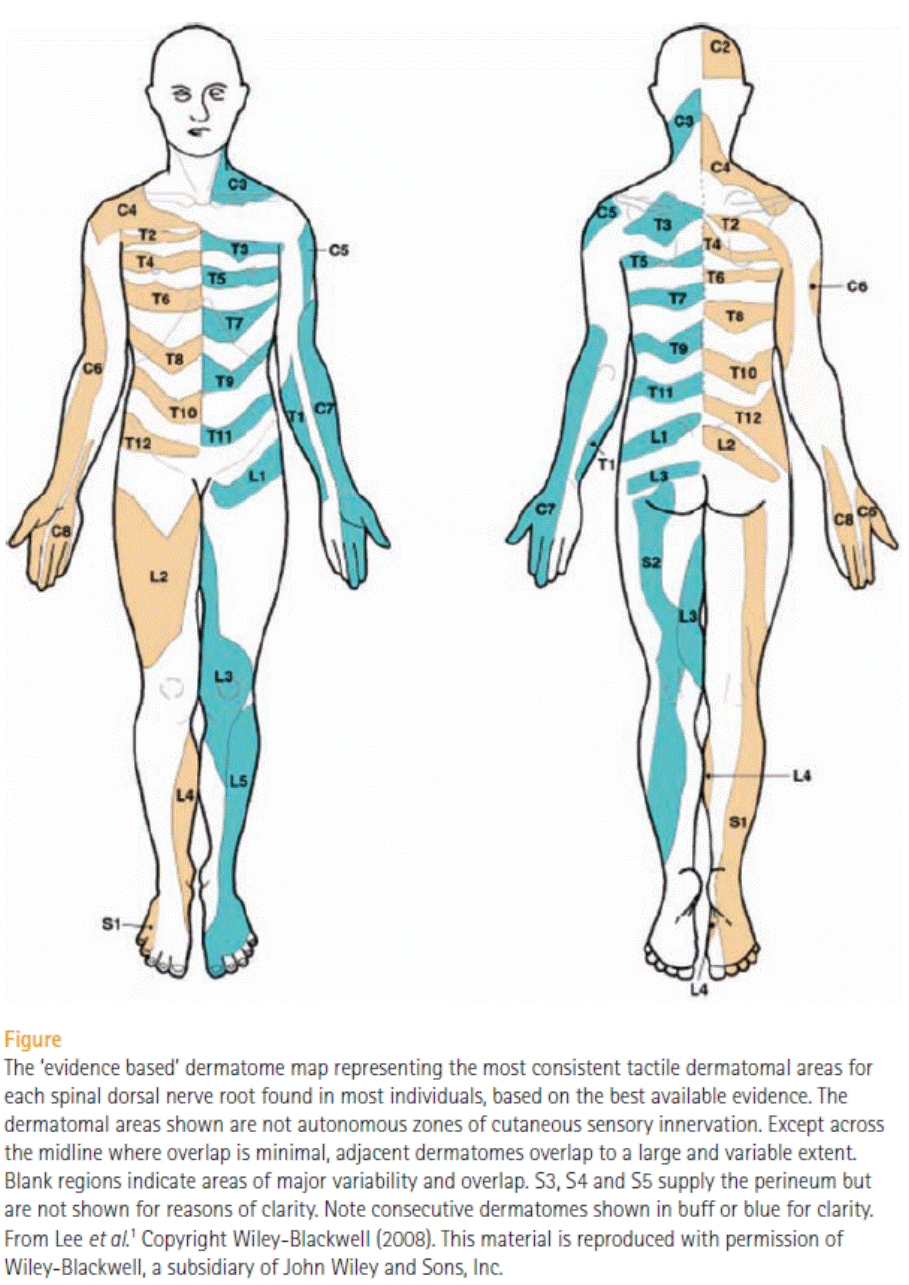
Sensation
|
Head |
|
|
C2 |
Back
of scalp |
|
C3 |
Supraclavicular
fossa |
|
|
|
|
Upper limb |
|
|
C4 |
Tip
of clavicle |
|
C5 |
Lateral
cubital fossa (just
proximal) |
|
C6 |
Thumb |
|
C7 |
Middle
finger |
|
C8 |
Little
finger |
|
T1 |
Medial
cubital fossa (just
distal) |
|
Trunk |
|
|
T4 |
Nipple |
|
T10 |
Umbilicus |
|
|
|
|
Lower Limb |
|
|
L2 |
Mid
anterior thigh |
|
L3 |
Medial
femoral condyle |
|
L4 |
Medial
malleolus |
|
L5 |
Dorsum
of 2nd/3rd MTPJ |
|
S1 |
Lateral
malleolus/heel |
|
S2 |
Popliteal
fossa |
|
S3 |
Ischial
tuberosity |
|
|
|
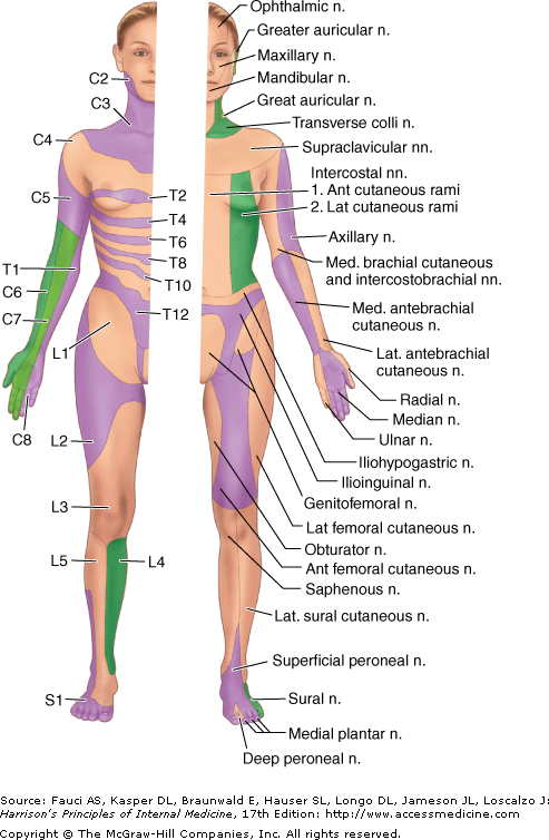
Upper
arm sensory
·
Lateral
- Terminal branch of
MSC nerve
- Medial cutaneous nerve of forearm
- Arises directly from
brachial plexus
- Posterior cutaneous nerver
of the arm
- Branch of the radial
nerve
Sensory
map
Rhomberg’s
test
·
Worsening of balance after eyes closed – requires 60sec technically
- Three elements feed into balance:
- Visual input
- Vestibular function
- Proprioception
- If the patient is unsteady before
closing eyes
- Cerebellar
dysfunction
- Vestibular function
- If the patient gets unsteady after the
eyes are closed
- Then likely
proprioceptive failure as vestibular function not adequate to maintain
posture on its own
Gait
Elements of the gait assessment
Posture
• Trunk – stooped vs upright
• Postural rexlexes – pull test
• Stance – narrow vs wide
Walking
• Initiation – hesitation, shuffling, magnetic
• Stepping
o Rhythm/Cadence (regular, irregular)
o Length (normal, short
o Trajectory (shallow, high-stepping)
o Speed
• Associated movements
o Trunk – sway (as in trendelenberg)
o Arm swing
Special manoeuvres
• Heel-toe
• Romberg’s test
• Walking backwards or running
Gait patterns
|
Gait |
Description |
Cause |
Cadence/Rhythm |
Step length |
Base |
|
(Spastic) Hemiparetic gait |
One
leg held stiffly and follows an arc |
Hemiplegia |
Slow |
Short |
Narrow |
|
Spastic (paraparetic) gait |
As
above however bilateral |
‘New’
onset paraparesis |
|
|
|
|
Scissoring gait |
As
above however tendency to adduction of
legs as well Short,
slow steps as if wading through water |
CP,
hereditary spastic paraplegia |
|
|
|
|
Parkinsons |
Shuffling
gait with reduced arm swing |
Parkinson’s
|
Slow
but can festinate |
Short |
Normal |
|
Apraxic/Prefrontal Marche a petits pas |
Similar
to parkinsons with wider base Feet
appear glued to floor (‘magnetic feet’), difficulty initiating and turning. |
Lacunar
infarcts NPH |
Slow |
Short |
Slightly
wide |
|
Waddling gait |
Swinging
shoulders from side to side. Lifting foot with help of trunk movt rather than hip adduction. |
Proximal
myopathy Muscular
dystrophy Hip
pathology – OA or congenital dislocations. |
Normal |
Normal |
Slightly
wide |
|
High stepping – unilateral |
High
step, foot hangs down. |
Foot
drop |
|
|
|
|
High stepping – bilateral (Steppage gait) |
As
above. Feet may slap the ground |
Bilateral
foot drop Peripheral
neuropathy – CMT MND |
Normal |
Normal |
Normal |
|
High stepping, broad based gait (Sensory ataxic) |
High
stepping, no foot drop, clumsy slapping down of feet, board base. Searching, patient watching feet. |
Posterior
column lesion – B12 or other sensory neuropathy MS Spinocerebellar
degeneration. |
Normal |
Short
|
Often
only slightly wide |
|
Cerebellar ataxia - truncal |
Loss
of truncal balance, increased body sway, disequilibrium Wide
based |
Midline
cerebellar structures |
Irregular,
overall often normal speed |
Slightly
short |
Wide |
|
Cerebellar ataxia - peripheral |
Irregular
steps with variable timing, length and direction. Fall to side of
lesion. If midline is involved as well
there may also be truncal imbalance |
Cerebellar
lobes |
|
|
|
|
Spastic Ataxia “bouncing gait” |
The
combination of increased tone, clonus and ataxia result in very unsteady gait
bouncing from one leg to the other and |
|
|
|
|
|
Dystonic |
Twisting,
athetoid or dystonic movements disrupt the gait May
be task specific – e.g. may disappear when walking backwards or running |
Dystonia |
Slow |
Normal |
Erratic |
|
|
|
|
Wide
based gait |
Sensitive
for a neurological disease, but not specific Atypical
parkinsonism Cerebellar,
sensory or vestibular ataxia Higher
level gait disorders NPH Functional |
|
Normal
base |
PD |
|
Narrow
base |
Parkinson’s
disease* Spastic
paraparesis |
|
Very
narrow/scissoring |
Spastic
paraparesis Huntington’s
disease (due to chorea) Functional |
|
Anterocollis/head drop |
MSA Myasthenia MND Polymyositis Focal
posterior cervical myositis |
|
Retrocollis |
PSP Cervical
dystonia Young
onset PD Drug
induced dystonia |
*
Patients with PD maintain good mediolateral stability late into the
disease. Remain able to ride a bicycle
late into disease.
Reference
for this table: Nonnekes et al. Neurological
disorders of gait, balance and posture.
Nat Rev Neurol 2018
Weakness
|
UMN |
Ischaemic,
focal lesion, vasculitis |
|
Anterior
horn cells |
SMA,
Lead, ALS, Poliomyelitis, Paraneoplastic
- |
|
Spinal
root |
Should
cause weakness AND matching sensory loss |
|
Peripheral
nerve |
GBS,
Leprosy, Myeloma, Amyloid, DM, Lead |
|
NM
Junction |
MG,
LEMS, Botulism, Organophosphate |
|
Muscle |
Polymyositis,
dermatomyositis, steroid, thyroid, hypoglycaemia, HIV, Muscular dystrophy. |
|
|
|
|
|
|
Approach to
Weakness