NCS
and EMG
NCS
EMG
Fibrillation
and positive sharp waves
Nerve
Conduction Studies
Definitions
• CMAP (Compound Muscle Action Potential)
o Summated voltage of individual muscle fibre action potentials
• SNAP
o Sensory nerve action potential
• Motor latency
- Distal
(terminal) latency – time to onset of CMAP
- Peak latency
– time to peak of CMAP
• Conduction velocity
o Calculated by difference in time between 2 different stimuli divided by the distance between them
• F-wave
o Signal travels back up motor nerve (antidromically) and activates some anterior horn cells producing a second small motor potential.
o Indicative of proximal pathology
• H-wave
o Low intensity stimulation (usually of tibial nerve) activates muscle spindles and fires reflex arc.
o May be more sensitive for mild neuropathies, S1 radiculopathy
• Dispersion
o ‘Spread’ of responses
o Widens in demyelination because some fibres are conducting slow and some fast.
• Conduction block
o Decrease in size of response by >20% between proximal and distal measurement points.
Late responses
F-Response
• Derived from “foot” where it was first recorded
• Antidromic impulse from stimulation travels back up to anterior horn cell
• A small (1-5%) and random population of anterior horn cells back-fire
• There is no synapse in the response
• Minimum F response latency measures the fastest conducting fibres – best measure
• F-wave persistence – usually >80%, lower limit of normal 50% (except peroneal)
• Chronodispersion – difference between slowest and fastest F responses – up to 4ms in upper limbs and 6ms in lower limbs
• F-responses may be absent in sleeping or sedated patients
• Reinforcement manoeuvres can be used to increase responses
• Normal values based on obtaining 10 responses
• Useful for:
o Polyradiculopathy – GBS
o Radiculopathy – limited to certain roots
- Median/Ulnar – C8-T1
- Peroneal/tibial - L5-S1
- Because early compressive radiculopathy might be expected to cause focal, predominantly sensory demyelination F-waves will be normal. Only a radiculopathy with significant injury to a large proportion of the motor fibres would be expected to decrease response.
- If there is a severe C8 radiculopathy the F-response might still be normal due to the T1 component (and similarly in the lower limb)
H-Reflex
• Named after Paul Hoffman who developed the test in 1918
• Can be elicited from many motor nerves before age 2 – after that age – only tibial
• True reflex
o Afferent – Ia muscle spindle
o Synapse
o Efferent – alpha motor neuron
• Evoked by low amplitude, long (1ms) stimulus
• Procedure
o Recording electrode just under ¾ of the way down from popliteal fossa to Achilles tendon (2-3 FB below point where gastroc meets soleus).
o Stimulate popliteal fossa with cathode placed proximally
o Low intensity, 2sec between stimuli
o If response cannot be obtained try – slightly plantarflexing ankle or Jendrassik manoeuvre
o As stimulus increases H reflex increases in amplitude and decreases in latency
o At higher stimulus an M-reponse appears, this also travels up the motor fibre, collides with and reduces the H-response
o Take the H-reponse with the shortest latency and measure maximal amplitude
• Normal values
o <34ms (varies according to height and age, use nomogram)
o <1.5ms difference between sides
o H/M amplitude ratio <50%
• Major uses:
o Early polyneuropathy
o S1 radiculopathy
o Early GBS
o Tibial and sciatic neuropathy, sacral plexopathy
Test
• Nerves often used
• Sensory and motor
• Median, ulnar, radial
• Sensory
• Sural, superficial peroneal, facial and accessory
• Motor
• Peroneal and tibial
•
Interpretation
|
|
Demyelination |
Axonal |
|
CMAP amplitude |
Normal |
Small |
|
Conduction block |
Present |
Non-present |
|
Distal motor latency |
Prolonged |
Normal/slightly prolonged |
|
Motor conduction velocity |
Reduced |
Normal/slightly reduced |
|
Sensory response |
Small absent |
Small absent |
|
F-wave |
Prolonged |
Normal/slightly prolonged |
|
|
|
|
EMG
Summary
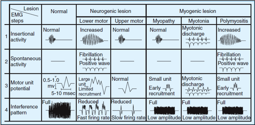
Insertional activity
• Burst of discharge on insertion of the needle
• Muscle fibres responding to structural damage/deformation from needle
• Decreased or absent – inexcitable fibres
o Fibrotic/atrophic muscle
o Familial periodic paralysis
• Prolonged – hyperexcitable fibres
o Denervation
o Myotonic disorder
o Myositis
Spontaneous activity
- Activity
occurring when the muscle is at rest, activity over which the patient has
no control
- Normal muscle
should be electrically silent
Endplate noise
• Low amplitude, monophasic, negative potentials
• Motor end plate potentials
• Rate: 20-40Hz
• Sound: Seashell
• Cause: Normal endplate
Endplate Spikes
• Source: single muscle fibre
o Firing due to irritation of terminal axon twigs by needle
• Initial negative deflection (c.f. fibrillations)
• Pattern: Irregular (sputtering)
• Rate: 5-50Hz
• Sound: sputtering, like fate in frying pan.
Fibrillation and positive sharp waves
• Are the same thing - Fibrillation – upwards deflections, PSWs – downwards deflections.
• Source: result from spontaneous firing of individual muscle fibres.
• Myopathies - if there is fibre splitting, part of the fibre becomes functionally denervated
• Occur as fibres become hyper-excitable when they lose their nerve supply and overexpress Ach receptors.
• Causes:
o Denervation
o Some muscle disease – esp. inflammatory myopathies
o Severe NMJ disease – e.g. botulism (rarely)
• Fibrillation
o Brief initial positive spike, then brief negative
o 1-5ms
o Low amplitude 10-100uV
o Very Regular 0.5-10Hz – may slow (c.f. endplate spikes – very irregular)
o Get smaller with more chronic denervation
o Sound – rain on roof
• Positive sharp waves
o Brief initial positive, long negative
o Low amplitude 10-100uV
o Caused by needle deforming muscle fibre
o Occasional discharges in distal muscles acceptable as normal
• Grading
o 0 - None
o +1 – Single trains in at least 2 areas
o +2 – Moderate number in 3 or more areas
o +3 – Many in all areas
o +4 – full interference pattern
Complex repetitive discharge
• Source: Depolarisation of single muscle fibre, followed by ephaptic spread to adjacent fibres, generates circuit
• Sound: Machine
• Rate: High frequency 5-100Hz
• Pattern: Perfectly regular, unless overdriven
• Usually identical each time
• Causes:
o Denervation – usually chronic rather than acute, such that group atrophy has occurred to allow denervated fibres to lie next to each other
o Some acute muscle disease with denervation
Myotonic discharges
• Source: Single muscle fibre
• Pattern:
o Waxing and waning of frequency and amplitude
• Rate: 20-150Hz
• Sound: Revving engine, Dive bomber
• Causes:
o Myotonic dystrophy
o Myotonia congenita
o Paramyotonia congenita
o Other myopathies: Acid maltase, polymyositis, myotubular myopathy
o Hyperkalaemic periodic paralysis
o Denervation (rarely)
Fasciculation
• Source: spontaneous firing of part or whole of motor unit
• Probably arise in the terminal braches of the motor nerve
• Morphology of normal CMAP (or abnormal if there is reinnervation)
• Rate:
o Often very slow (1-2Hz – c.f. minimum 4-5Hz of voluntary activity)
o Overall range 0.1-10Hz
• Pattern: Irregular
• Sound: Corn popping
• Causes: Occur in neurogenic conditions
Doublets/Multiplets
• Grouped fasciculations
• Same significance as fasciculations except are characteristically seen in hypocalcaemia
Myokymia
• Grouped fasciculations
• Rhythmic grouped spontaneous firing of the same motor unit
• Spontaneous depolarisation or ephaptic transmission along demyelinated segments of nerves
• Rate:
o Intraburst 5-60Hz
o Interburst 1-5Hz
• Sound: Marching soldiers
• Causes:
o Radiation injury (usually brachial plexus)
o GBS (Facial)
o MS (facial)
o Pontine tumours (Facial)
o Hypocalcaemia
o Timber rattlesnake venom
o Rarely also seen in:
- GBS (limbs)
- CIDP
- Nerve entrapments
- Radiculopathy
Cramps
• Painful, involuntary contractions of muscle that tend to occur when muscle is in shortened position and contracting
• Source: High frequency discharges of motor axons/motor units
• Rate: 40-75Hz
Neuromyotonic discharges
• Involuntary spontaneous discharge of motor units and their axons
• Rate: Very high frequency 150-250Hz
• Pattern: Decrementing, repetitive discharge of a single motor unit
• Sound: Pinging
• Cause:
o Neuomyotonic syndromes (Isaac’s syndrome etc)
o Extremely chronic neuropathic disease (e.g. polio and SMA)
o Hereditary neuromyotonia
Voluntary Activity
• Analyse:
o Morphology
- Duration
- Polyphasia
- Amplitude
o Stability
o Firing Pattern
- Activation
- Recruitment
- Interference
Morphology
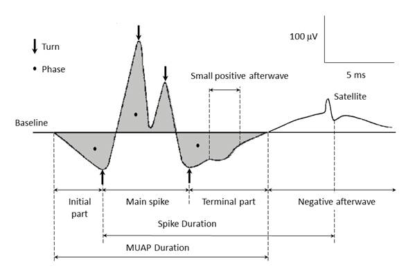
Duration
• Parameter that best reflects number of muscle fibres in motor unit
• Typical 5-15ms
• Increased with age
• Decreases with increased temperature
• Increased in distal muscles
• Sound – duration is inversely proportional to pitch (dull to crisp)
Polyphasia
• Measure of synchrony
• Nonspecific
• Normal:
o 2-4phases
o <5-10% of MUAPs polyphasic
o Except deltoid: normal <25%
• Sound: high frequency clicking
Amplitude
• Measured from negative to positive peak
• Normal 0.1-2mV
• Increased size with motor unit synchrony
• Sound: proportional to volume
Stability
• Measure of the similarity of the morphology of an individual MUAP each time it fires
• Lack of stability correlates with impaired NMJ function
Firing Pattern
Activation
• Ability to increase firing rate
• Causes:
o Central disease – CNS – MS/stroke
o Reluctance to activate due to pain
Recruitment
• Ability to recruit more MUAPs as the firing rate and force increase
• First motor unit starts with rate of 5Hz, by 10Hz a second unite should activate and so on.
• 5:1 ratio
• Decreased recruitment
o Loss of MUAP axons
o Conduction block
Early recruitment
• Too many units firing for the degree of force being generated (examiner needs to be judging amount of force)
• Seen when there is dropout of individual muscle fibres (Myopathies/NMJ blockade)
• Each MUAP generates less force, therefore more units needed early
Common
patterns
|
|
EMG |
NCS |
|||||||||||
|
|
MUAP Morphology |
MUAP Firing
Pattern |
Spontaneous |
Motor |
Sensory |
||||||||
|
|
Dur. |
Amp. |
Pha. |
Act. |
Recr. |
Fibs/PSW |
Amp |
DL |
CV |
F-M |
Amp |
DL |
CV |
|
Neuropathic-axonal |
|
|
|
|
|
|
|
|
|
|
|
|
|
|
Acute (several
days – few weeks) |
N |
N |
N |
N |
D |
N |
D |
N/I |
N/D |
N/I |
D |
N/I |
N/D |
|
Subacute (several
weeks) |
N |
N |
N |
N |
D |
I++ |
D |
N/I |
N/D |
N/I |
D |
N/I |
N/D |
|
Chronic
(inactive: months-yrs) |
I |
I |
I |
N |
D |
N |
N/D |
N/I |
N/D |
N/I |
N/D |
N/I |
N/D |
|
Neuropathic-demyelinating |
|
|
|
|
|
|
|
|
|
|
|
|
|
|
Proximal lesion -
slowing |
N |
N |
N |
N |
N |
N |
N |
N |
Distal-N Prox- D |
I++ |
N |
N |
N |
|
Proximal lesion –
with block |
N |
N |
N |
N |
D |
N |
N |
N |
Distal-N Prox- D |
I++ |
N |
N |
N |
|
Distal Lesion –
slowing |
N |
N |
N |
N |
N |
N |
N |
I |
D |
N |
N/D |
I++ |
D++ |
|
Distal lesion -
block |
N |
N |
N |
N |
D |
N |
D |
I |
D |
N |
D |
I++ |
D++ |
|
Early
reinnervation after severe denervation |
D |
D |
I |
N |
D++ |
|
D++ |
N/I |
N/D |
N/I |
D++ |
N/D |
N/I |
|
Myopathic |
|
|
|
|
|
|
|
|
|
|
|
|
|
|
Acute |
D |
D |
I |
N |
N/Early |
N/+/Myotonia |
N/D |
N |
N |
N |
N |
N |
N |
|
Chronic (with
denervating features) |
D/I |
D/I |
I |
N |
N/Early |
N/+/CRDs |
N/D |
N |
N |
N |
N |
N |
N |
|
Endstage |
D/I |
D/I |
I |
N |
D++ |
|
N/D |
N |
N |
N |
N |
N |
N |
|
NMJ disorders |
|
|
|
|
|
|
|
|
|
|
|
|
|
|
Increased jitter |
N |
N |
N |
N |
N |
N |
PreS-N PostS-D |
N |
N |
N |
N |
N |
N |
|
Intermittent
block |
N/D |
N/D |
N/I |
N |
N/Early |
I+ |
|||||||
|
Severe block |
D |
D |
I |
N |
D++ |
I++ |
|||||||
|
|
|
|
|
|
|
|
|
|
|
|
|
|
|
|
CNS disorders |
N |
N |
N |
D++ |
N |
|
|
|
|
|
|
|
|
Treatment
Patterns
of Nerve Injury
Time related changes
Axonal lesions
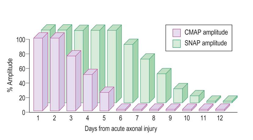
• Initially distally stimulated nerves conduct normally, until Wallerian degeneration takes place (as per graph).
• Abnormal spontaneous activity – time to develop depends on distance from lesion to muscle. E.g.
o L5-S1 nerve root lesion fibs and PSWs develop after:
- 10-14days - Paraspinal muscles
- 2-3 weeks - Proximal thigh
- 3-4 weeks - Leg
- 5-6 weeks – distal leg/foot
o If it is a distal lesion, near NMJ, then may only take a few days to develop.
|
|
Immediate |
Hyperacute <3 days |
Acute >1Wk to <3-6 Wk |
Subacute >1 Wk to < 3-6 Wk |
Subacute/Chronic >2-3 Months to Many months |
Chronic Years |
|
Clinical findings |
Abnormal |
Abnormal |
Abnormal |
Abnormal |
Abnormal |
Abnormal/Normal |
|
NCS |
Normal |
Normal |
Abnormal |
Abnormal |
Abnormal |
Abnormal/Normal |
|
MUAP recruitment |
Decreased |
Decreased |
Decreased |
Decreased |
Decreased |
Decreased |
|
Spontaneous activity |
Normal |
Normal |
Normal/ abnormal |
Abnormal |
Abnormal |
Normal |
|
MUAP morphology |
Normal |
Normal |
Normal |
Normal |
Reinnervated |
Reinnervated |