Facial Muscles |
|||||
|
Muscle |
Nerve |
Origin |
Insertion |
Action |
Needle |
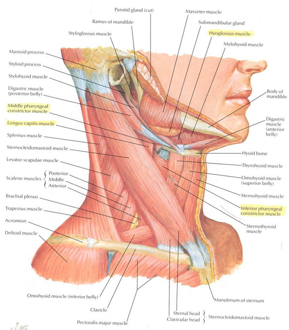
Oral cavity |
|||||
|
Genioglossus |
Hypoglossal |
|
|
Major
extrinsic muscle of the tongue – protrudes tongue. |
Insert
from beneath the anterior mandible, just lateral to the midline. |
|
|
|
|
|
|
|
Muscles of mastication |
|||||
|
Masseter |
TGN
V3 Mandibular division |
Zygomatic
arch |
Angle
of mandible |
Elevates
and protrudes mandible |
2
FB ant. To angle of jaw and 1-2 FB cephalad. |
|
Temporalis |
Temporal
foss |
Coronoid
process of mandible |
Elevates
and retracts mandible |
|
|
|
Lateral
pterygoid |
Sphenoid
bone |
Head
of mandible (near TMJ) |
Initiates
mouth opening by protruding mandible |
|
|
|
Medial
pterygoid |
Sphenoid
bone |
Angle
of mandible (on interior surface) |
Elevates
the mandible |
|
|
Forehead |
|||||
|
Frontalis |
Facial
nerve, frontal branch |
|
|
|
2
FB above the middle of the eyebrow |
|
Orbital opening/Palpebral fissure |
|||||
|
Orbicularis
oculi |
Facial
nerve, temporal branch |
Medial
orbit |
Skin
around margin of orbit |
Closes
eyelids |
Lat.
inferior ridge of eye socket |
|
Corrugator
supercilii |
|
|
|
Wrinkles
the eyebrow – Frown Thoughtful
brow |
|
|
Depressor
supercilli |
|
|
|
Lowers
the eyebrow |
|
|
|
|
|
|
|
|
Nose |
|||||
|
Nasalis |
|
Maxilla |
Nasal
cartlidges |
Narrows
the nose |
|
|
Procerus |
|
Continuation
of frontalis |
Superior
nose |
Wrinkles
root of nose (by pulling nose up and eyebrows down) |
|
|
Levator labii superioris
alaeque nasi |
|
|
|
Elevates
upper lip and nasal alae Disapproval |
|
Mouth |
|
|
|
|
|
|
Orbicularis
oris |
|
|
|
Closes
the mouth (and protrudes/purses lips) |
|
|
Buccinator |
|
|
|
Muscle
of the cheek – presses check against teeth – aids in eating. Satisfaction |
|
|
Zygomaticus
major |
|
Zygomatic
arch |
Corner
of mouth |
Smiling |
|
|
|
|
|
|
|
|
|
Risorius |
|
Platysma |
Corner
of mouth |
Draws
sides of mouth laterally Purposeful
action |
|
|
Levator labii superioris |
|
Maxilla |
Upper
lip |
Elevates
lip |
|
|
Levator anguli
oris |
|
|
|
Pulls
the corner of the mouth upwards Self-satisfaction |
|
|
Depressor
anguli oris |
|
|
|
Pulls
corners of mouth down Sadness |
|
|
Depressor
labii inferioris |
|
|
|
Pulls
lower lip down Perseverence |
|
|
Mentalis |
Facial
nerve, mandibular branch |
Mandible |
Skin
of chin |
Pulls
the skin on the chin upwards – elevating and protruding the lower lip |
Insert
into chin, have patient purse lips |
|
|
|
|
|
|
|
Neck |
|||||
|
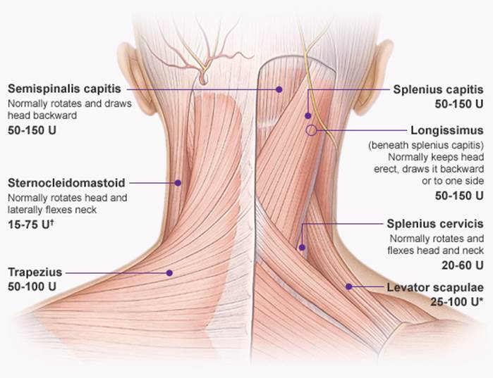
Sterocleido-mastoid |
|
|
|
|
|
|
Splenius
capitus |
Middle
cervical spinal nerves |
Spinous
processes of superior 6 thoracic vertebrae |
Mastoid
process and lateral 1/3 or superior nuchal line |
Laterally
flexes and rotates the head to the same side |
|
|
Posterior
scalene |
Spinal
nerves C7 and C8 |
Transverse
processes of C4-C6 vertebrae |
External
border of 2nd rib |
Flexes
neck laterally, elevates 2nd rib |
|
|
Middle
scalene |
|
|
Sup.
Surface of 1st rib |
Flexes
neck laterally, elevates 1st rib |
|
|
Anterior
scalene |
Spinal
nerves C4, C5, C6 |
|
1st
rib |
Elevates
1st rib, laterally rotates neck |
|
|
Semispinalis
capitus |
|
Transverse
processes of thoracic spine |
Occipital
bone |
Neck
extension |
|
Upper limb |
|||||||||||||||||
Shoulder Gridle |
|||||||||||||||||
|
Muscle |
Nerve |
Trunk |
Division |
Cord |
C1 |
C2 |
C3 |
C4 |
C5 |
C6 |
C7 |
C8 |
T1 |
Origin |
Insertion |
Action |
Needle |
|
Trapezius |
Accessory |
Cranial
nerve |
|
|
|
|
|
|
|
|
|
Nuchal
ligament, spinous processes C7-C12 |
Clavicle,
acromion, spine of scapula |
Elevates,
retracts and rotates scapula |
At
point where shoulder meets the neck. |
||
|
Rhomboids |
Dorsal
Scapular |
Off
C5 nerve root |
|
|
|
|
|
|
|
|
|
Spinous
processes C7-T5 |
Medial
border of scapular – from spine down. |
Retracts
scapula and rotates the glenoid cavity down.
Fixes scapula to chest wall. |
Mid-point
b/n medial border of scapula and spine.
Ask patient to brace shoulder backwards |
||
|
Levator Scapulae |
Dorsal
Scapular |
Off
C4 and C5 nerve root |
|
|
|
|
|
|
|
|
|
Transverse
processes C1-C4 |
Superior,
medial scapular |
Elevates
scapula and tilts its glenoid cavity inferiorly by rotating scapula |
|
||
|
Serratus
Anterior |
Long
thoracic |
Off
C5, C6, C7 |
|
|
|
|
|
|
|
|
|
Superior
8 or 9 ribs |
Costal
aspect of medial scapular |
Protracts
and stabilizes scapula, assists in upward rotation. |
Over
6th rib, mid-axillary line. |
||
|
Latissimus
dorsi (C6, C7, C8) |
Thoracodorsal |
All
3 |
Posterior |
Posterior |
|
|
|
|
|
|
|
|
|
Spinous
processes from T7 down |
Medial,
proximal humerus |
Extends,
adducts and medially rotates shoulder |
Lat.
to tip of scapula at post. Axillary line. Have the pt. extend shoulder while
adducted. |
|
Supraspinatus |
Suprascapular
|
Upper |
|
|
|
|
|
|
|
|
|
|
|
Supraspinous
fossa of scapula |
superior
facet of greater tubercle of humerus |
Abduction
of arm (first 30deg) Stablisation of humerus |
Just
sup. And med. To mid-point of scapular spine. |
|
Infraspinatus |
Suprascapular
|
Upper |
|
|
|
|
|
|
|
|
|
|
|
Infraspinous
fossa of the scapula |
middle
facet of greater tubercle of the humerus |
Lateral
rotation of arm and stabilizes humerus |
1-2FB
below mid-point of scapular spine. |
|
Subscapularus |
Upper/lower
subscapular |
Upper |
Posterior |
Posterior |
|
|
|
|
|
|
|
|
|
Subscapular
fossa |
Lesser
tubercle of humerus |
Internally
(medially) rotates humerus; stabilizes shoulder |
|
|
Teres
major |
Lower
subscapular |
Upper |
Posterior |
Posterior |
|
|
|
|
|
|
|
|
|
Posterior
aspect of the inferior angle of the scapula |
Medial
lip intertubercular
sulcus of the humerus |
Internal
rotation (medial rotation) of the humerus |
|
|
Teres
minor |
Axillary |
Upper |
Posterior |
Posterior |
|
|
|
|
|
|
|
|
|
lateral
border of the scapula |
inferior
facet of greater tubercle of the humerus |
Externally
rotates arm, stabilises humerus |
2/3
along line from inf. Point of scapular to acromium.
|
|
Deltoid |
Axillary |
Upper |
Posterior |
Posterior |
|
|
|
|
|
|
|
|
|
Lateral
third of the clavicle, acromion, spine of the scapula |
Deltoid
tuberosity of humerus |
shoulder
abduction, flexion and extension |
4cm
distal to acromium in midpoint. |
|
Pectoralis
major |
Lateral,
medial pectoral |
All
three |
Anterior |
Lateral,
medial |
|
|
|
|
|
|
|
|
|
Medial
half of the clavicle, anterior surface of the sternum, the superior six
costal cartilages |
bicipital
groove of the humerus |
As a whole, adducts and medially
rotates the humerus. It also draws the scapula anteriorly and inferiorly. |
Ant.
Inf shoulder (5 FB below humeral head) Have
pt abduct shoulder. |
|
Pectoralis
minor |
Lateral,
medial pectoral |
All
three |
Anterior |
Lateral |
|
|
|
|
|
|
|
|
|
Third
to fifth ribs, near their costal cartilages |
Medial
border and superior surface of the coracoid process of the scapula |
Stabilizes
the scapula by drawing it inferiorly and anteriorly against the thoracic wall |
|
|
Muscle |
Nerve |
Trunk |
Cord |
Division |
C1 |
C2 |
C3 |
C4 |
C5 |
C6 |
C7 |
C8 |
T1 |
Origin |
Insertion |
Action |
|
|
Biceps
brachii |
Musc. |
Lateral |
Anterior |
Lateral |
|
|
|
|
|
|
|
|
|
Short
head: coracoid process of the scapula. Long
head: supraglenoid tubercle |
Radial
tuberosity and bicipital aponeurosis into deep fascia on medial part of
forearm |
Flexes
elbow (when supine) and supinates forearm |
Midpoint
b/n biceps tendon and shoulder |
|
Brachialis |
Musc. |
Lateral |
Anterior |
Lateral |
|
|
|
|
|
|
|
|
|
Distal
anterior humerus |
Coronoid
process and tuberosity of ulna |
Flexes
forearm (in all positions) |
|
Median |
|||||||||||||||||
|
Muscle |
Nerve |
Trunk |
Division |
Cord |
C1 |
C2 |
C3 |
C4 |
C5 |
C6 |
C7 |
C8 |
T1 |
Origin |
Insertion |
Action |
Needle |
|
Pronator
teres (PT) |
Median |
Upper,
Middle |
Anterior |
Lateral |
|
|
|
|
|
|
|
|
|
Medial
epicondyle of the humerus (+some
radial and ulna) |
Mid
lateral radius |
Pronates
and flexes forearm |
2
FB distal to mid-point b/n biceps tendon and med. Epicondyle. |
|
Flexor
carpi radialis (FCR) |
Median |
Upper,
Middle |
Anterior |
Lateral |
|
|
|
|
|
|
|
|
|
Base
of 2nd Metacarpal |
Flexes
and abducts hand at wrist |
4
FB distal to mid-point b/n biceps tendon and med. Epicondyle. On line to mid point of wrist. |
|
|
Palmaris
longus |
Median |
|
|
|
|
|
|
|
|
|
|
|
|
Flexor
retinaculum and palma aponeurosis |
Flexes
hand and tightens palmar aponeurosis |
|
|
|
Flexor
digitorum superficialis (FDS) |
Median |
Middle,
Lower |
Anterior |
Lateral,
Medial |
|
|
|
|
|
|
* |
* |
|
Middle
phalanges |
Flexes
middle phalanges at PIP and Proximal phalanges at MCP joints |
Just
med. To mid-point b/n biceps tendon and mid wrist. |
|
|
Flexor
digitorum profundus (II and III) (FDP) |
Median
(AIN) |
Middle,
Lower |
Anterior |
Lateral,
Medial |
|
|
|
|
|
|
* |
|
|
Proximal
Ulna & interosseous membrane |
Distal
phalanges |
Flexes
distal phalanges at DIP |
3-4
FB distal to olecranon, on ulnar side of forearm (deep) |
|
Flexor
pollicis longus (FPL) |
Median
(AIN) |
Middle,
Lower |
Anterior |
Lateral,
Medial |
|
|
|
|
|
|
* |
|
|
Radius
and interosseous membrane |
Base
of distal phalanx of thumb |
Flexes
phalanges of thumb (1st digit) |
Over
radius 1/3 of way up to elbow. |
|
Pronator
quadratus (PQ) |
Median (AIN) |
Middle,
Lower |
Anterior |
Lateral,
Medial |
|
|
|
|
|
|
* |
|
|
Distal
anterior ulna |
Distal
anterior radius |
Pronates
forearm |
Arm
half way supinated.
Dorsal arm, 3 Fb proximal to styloids at
mid-point. – deep. |
|
Lumbricals
I, II |
Median
(RB) |
Lower |
Anterior |
Medial |
|
|
|
|
|
|
|
|
|
FDS
tendon in palm |
Lateral
prox. phalanx (ext. expansion) |
Flex
MCP and extend PIPs |
|
|
Abductor
pollicis brevis (ABP) |
Median
(RB) |
Lower |
Anterior |
Medial |
|
|
|
|
|
|
|
|
|
Flex
ret. Scaphoid, trapezium |
Lateral
side of base of prox. phalanx |
Abduct
and oppose thumb |
Lat.
thenar eminence |
|
Flexor
pollicis brevis |
Median
(RB) (+/-
ulnar) |
Lower |
Anterior |
Medial |
|
|
|
|
|
|
|
|
|
Med-lateral
side of base of prox. phalanx |
Flexes
thumb |
Med.
To midpoint of thenar eminence |
|
|
Opponens pollicis |
Median
(RB) |
Lower |
Anterior |
Medial |
|
|
|
|
|
|
|
|
|
Lateral
1st MCP |
Opposes
thumb |
Lat.
thenar eminence, just ant. To 1st metacarpal. |
|
Ulnar |
|||||||||||||||||
|
Muscle |
Nerve |
Trunk |
Division |
Cord |
C1 |
C2 |
C3 |
C4 |
C5 |
C6 |
C7 |
C8 |
T1 |
Origin |
Insertion |
Action |
Needle |
|
Flexor
carpi ulnaris (FCU) |
Ulnar |
Lower |
Anterior |
Medial |
|
|
|
|
|
|
|
|
|
Medial
epicondyle of humerus |
Pisiform,
hamate, 5th MC. |
Flexes
and adducts wrist |
Med.
Forearm, mid point b/n elbow and wrist |
|
Flexor
digitorum profundus IV and V (FDP) |
Ulnar |
Lower |
Anterior |
Medial |
|
|
|
|
|
|
|
|
|
Proximal
Ulna and interosseous membrane |
Distal
phalanges |
Flexes
distal phalanges at DIP |
3-4
FB distal to olecranon, on ulnar side of forearm (superficial) |
|
Abductor
digiti minimi (ADM) |
Ulnar |
Lower |
Anterior |
Medial |
|
|
|
|
|
|
|
|
|
Pisiform |
Med.
Base, prox. phalanx little finger |
Adducts
digit 5 |
Med.
Hand, mid point of 5th metacarpal. |
|
Lumbricales III and IV |
Ulnar |
Lower |
Anterior |
Medial |
|
|
|
|
|
|
|
|
|
Tendons
of FDP |
Lat.
prox. phalanx |
Flex
MCPJ (and extend IPJs) |
|
|
Palmar
interossei |
Ulnar |
Lower |
Anterior |
Medial |
|
|
|
|
|
|
|
|
|
Palmar
surfaces 2nd, 4th 5th MC |
Base
of prox. phalanx |
Adducts
digits (aids flexion) |
|
|
First
dorsal interosseous (FDI) |
Ulnar |
Lower |
Anterior |
Medial |
|
|
|
|
|
|
|
|
|
Adjacent
metacarpals |
Base
prox phalanges |
Abduct
digits ( aids flexion) |
Dorsal
hand half way between 1st and 2nd
MCPJs. |
|
Adductor
pollicis |
Ulnar |
Lower |
Anterior |
Medial |
|
|
|
|
|
|
|
|
|
2nd
and 3rd metacarpals |
Med.
Base, prox. phalanx of thumb |
Adducts
thumb |
|
Radial |
|||||||||||||||||
|
Muscle |
Nerve |
Trunk |
Division |
Cord |
C1 |
C2 |
C3 |
C4 |
C5 |
C6 |
C7 |
C8 |
T1 |
Origin |
Insertion |
Action |
Needle |
|
Triceps |
Radial |
Upper,
Middle |
Posterior |
Posterior |
|
|
|
|
|
|
|
|
|
Humerus
and scapula |
Ulna
– olecranon |
Extends
forearm |
Mid point b/n lat. epicondyle and shoulder |
|
Brachioradialis (BR) |
Radial |
Upper |
|
|
|
|
|
|
|
|
|
Lateral
supracondylar ridge of humerus |
Lateral,
distal radius |
Flexes
forearm |
3-4
FB dis. To mid-point b/n biceps tendon and lat. epicondyle |
||
|
Anconeus |
|
|
|
|
|
|
|
|
|
|
|
|
|
|
2
FB distal to olecranon, just above the ulna |
||
|
Extensor
carpi radialis longus (ECRL) |
Radial |
Upper,
Middle |
|
|
|
|
|
|
* |
|
|
Lateral
supracondylar ridge of humerus |
Base
of 2nd metacarpal |
Extend
and abduct hand at wrist |
Just
above lat. epicondyle |
||
|
Extensor
carpi radialis brevis (ECRB) |
Radial |
|
|
|
|
|
|
|
|
|
|
|
|
|
|
||
|
Supinator |
Radial
- deep
branch |
Upper,
Middle |
|
|
|
|
|
|
|
|
|
Lateral
epicondyle of humerus + prox ulna |
Prox.
third of radius |
Supinates
forearm |
|
||
|
Extensor
carpi ulnaris (ECU) |
Radial
– Posterior. Interosseous. |
Middle,
Lower |
|
|
|
|
|
|
|
|
|
Base
of 5th metacarpal |
Extends
and adducts hand at wrist |
Mid point of ulnar on dorsal arm (slightly towards radius) |
|||
|
Extensor
digitorum (communis) |
Middle,
Lower |
|
|
|
|
|
|
|
|
|
Lateral
epicondyle of humerus |
Extensor
expansions of digits |
Extends
digits at MCP joints + extends hand at wrist |
3-4
FB distal to olecranon, 3 FB above the ulna |
|||
|
Abductor
pollicis longus |
Middle,
Lower |
|
|
|
|
|
|
|
|
|
Posterior
ulna |
Base of 1st
metacarpal |
Abducts
thumb and extends it at carpometacarpal joint |
|
|||
|
Extensor
pollicis longus |
Middle,
Lower |
|
|
|
|
|
|
|
|
|
Posterior
ulna |
Base
of distal phalanx of thumb |
Extends
distal phalanx of thumb at MCP and IP joints |
|
|||
|
Extensor
pollicis brevis |
|
|
|
|
|
|
|
|
|
|
|
|
|
|
|||
|
Extensor
indicis (EIP) |
Middle,
Lower |
|
|
|
|
|
|
|
|
|
Posterior
ulna |
Extensor
expansion of 2nd digit |
Extends
2nd digit and helps extend hand at wrist |
2
FB prox and slightly medial to ulnar styloid on
dorsum. |
|||
For
needle insertion anatomy see Muscle diagrams
Lower Limb |
|||||||||||
Femoral Nerve |
|||||||||||
|
Muscle |
Nerve |
L2 |
L3 |
L4 |
L5 |
S1 |
S2 |
Origin |
Insertion |
Action |
Needle |
|
Iliopsoas |
Femoral
/Lumbar plexus |
* |
|
|
|
|
|
Sides
of T12-L5 vertebrae +Iliac fossa |
Lesser
trochanter of femur |
Flex
thigh at the hip |
2-3
FB lat. to the femoral pulse and just below the inguinal ligament |
|
Sartorius |
Femoral |
|
|
* |
|
|
|
ASIS |
Superior,
medial tibia |
Flexes,
Abducts and laterally rotates thigh, flexes knee |
|
|
Rectus
femoris |
Femoral |
|
|
|
|
|
|
Ant.
Inf. Iliac Spine |
Patella |
Extend
leg at knee joint (Recutus femoris also flexes hip) |
Ant.
Thigh, mid-point b/n hip and knee. |
|
Vastus
lateralis |
Femoral |
|
|
|
|
|
|
Greater
trochanter |
Lat.
thigh, 4-5 FB proximal to lateral knee |
||
|
Vastus
intermedius |
Femoral |
|
|
|
|
|
|
Anterior
femur |
Deep
to vastus lat. and rectus fem. |
||
|
Vastus
medialis |
Femoral |
|
|
|
|
|
|
Medial
femur |
Med.
Thigh 3-4 FB prox. to the med. Knee. |
||
Obturator Nerve |
|||||||||||
|
Adductor
longus |
Obturator |
|
|
|
|
|
|
Pubis |
Middle
third of femur |
Adducts
thigh |
Medial
thigh 3-4 FB distal to the pubis. Hard
to differentiate the individual muscles – treat as a group. |
|
Gracillis |
Obturator |
|
|
|
|
|
|
Inferior
pubic ramus |
Superior,
medial tibia |
Adducts
thigh, flexes leg, medial rotation |
|
|
Adductor
magnus |
Obturator |
|
|
|
|
|
|
Ischial
tuberosity |
Medial
femur |
Adducts
and flexes thigh |
|
|
Adductor
brevis |
Obturator |
|
|
|
|
|
|
Inferior
pubic ramus |
Proximal
medial femur |
Adducts
and flexes thigh |
|
Gluteal Muscles
|
|||||||||||
|
Gluteus
maximus |
Inferior
gluteal |
|
|
|
|
|
|
Ilium,
sacrum, coccyx |
Iliotibial
tract and proximal femur |
Extends
thigh (esp. from flexed), lateral rotation |
Upper outer quadrant
of the buttock (have patient extend thigh or squeeze buttocks) |
|
Gluteus
medius |
Superior
gluteal |
|
|
|
|
|
|
Ilium,
proximal |
Lateral
greater trochanter |
Abduct
and medially rotate thigh, keep pelvis level when opposite foot is raised |
2-3
FB distal to midpoint of iliac crest (post. To line b/n ASIS and greater
trochanter) |
|
Gluteus
minimus |
Superior
gluteal |
|
|
|
|
|
|
Ilium
distal |
Anterior
greater trochanter |
|
|
|
Piriformis |
Spinal
roots |
|
|
|
|
|
|
Anterior
sacrum |
Superior
greater trochanter |
Lat.
rotate extended thigh, abduct flexed thigh |
|
|
Tensor
fascia lata |
Superior
gluteal |
|
|
|
|
|
|
ASIS |
Iliotibial
band (attaches to proximal tibia) |
Thigh
flexor, some medial rotation Tenses
iliotibial band to stabilise thigh on knee. |
2
cm lat. and 5cm distal to ASIS (just anterior to greater trochanter). |
Sciatic Nerve |
|||||||||||
|
Muscle |
Nerve |
L2 |
L3 |
L4 |
L5 |
S1 |
S2 |
Origin |
Insertion |
Action |
Needle |
|
Semimembranousus |
Sciatic
– tibial division |
|
|
* |
|
|
|
Ischial
tuberosity |
Medial,
superior tibia |
Extend
thigh, flex and medially rotate leg |
3-4
FB pox. To med. Knee, lat. to the tendon of semitendinous |
|
Semitendinosus |
|
|
* |
|
|
|
Mid point b/n med. Knee and ischial tuberosity. |
||||
|
Biceps
femoris |
Sciatic Long
head: Tibial division Short
Head: Common peroneal division |
|
|
|
|
|
|
Long
head: Ischial tuberosity Short
head: distal femur |
Fibula
head |
Extends
thigh, flexes and laterally rotates leg |
Short
head: 3-4 FB prox. to lat. knee.
Medial to the tendon of long head. Long
head: Mid point between lateral knee and ischial
tuberosity. |
Peroneal Nerve |
|||||||||||
|
Muscle |
Nerve |
L2 |
L3 |
L4 |
L5 |
S1 |
S2 |
Origin |
Insertion |
Action |
Needle |
|
Tibialis
anterior (TA) |
Deep
peroneal |
|
|
|
|
|
|
Proximal,
lateral tibia |
Medial
cuneiform and base 1st metacarpal |
Dorsiflexion
and foot inversion |
Just
lat. to tibial crest 2/3rd way up leg |
|
Extensor
digitorum longus |
Deep
peroneal |
|
|
|
|
* |
|
Proximal,
lateral, tibia and fibular |
Middle
and distal phalanges of lateral 4 digits |
Extends
lateral 4 digits and dorsiflexes ankle |
3-4
FB lat. to tbial crest, mid leg. B/n TA and PL. |
|
Exensor hallucis longus
(EHL) |
Deep
peroneal |
|
|
|
|
|
|
Mid
fibular |
Base
of distal phalanx of great toe |
Extends
great toe and dorsiflexes ankle |
|
|
Extensor
digitorum brevis (EDB) |
Deep
peroneal |
|
|
|
|
|
|
Dorsal
surface of calcaneus |
Proximal
dorsal region of middle phalanges 2, 3 and 4 (EHB
is an extension of the same muscle to 1st phalanges) |
Aids
extension of digits 2-4 (N.B digit 5 extended by EDL only) |
2-3
FB below lateral malleolus on dorsolateral foot |
|
Extensor
hallucis brevis (EHB) |
Deep
peroneal |
|
|
|
|
|
|
Aids
extension of great toe |
3-4
FB above ankle, just lat. to TA tendon |
||
|
Peroneus
longus |
Superficial
peroneal |
|
|
|
|
|
|
Superior,
lateral fibular |
Base
of 1st metatarsal (crosses under sole of foot) |
Evert
and weakly plantarflex the foot |
Lat.
calf, 3-4 FB below fibuar head |
|
Peroneus
brevis |
Superficial
peroneal |
|
|
|
|
|
|
Inferior,
lateral fibular |
Base
of 5th metatarsal |
|
|
Tibial Nerve
|
|||||||||||
|
Muscle |
Nerve |
L2 |
L3 |
L4 |
L5 |
S1 |
S2 |
Origin |
Insertion |
Action |
Needle |
|
Gastrocnemius
(medial) |
Tibial |
|
|
|
|
|
* |
Medial
condyle of femur |
Calcaneal
(Achilles tendon) |
Plantarfexion of foot Flexion
of knee |
Pos-med.
calf |
|
Gastrocnemius
(lateral) |
Tibial |
|
|
|
|
|
|
Lateral
condyle of femur |
|
|
|
|
Soleus |
Tibial |
|
|
|
|
|
|
Fibular
head, proximal tib and fib |
Plantarflexes
foot |
Medial
to the tibia, below half way point of leg, angle
back. |
|
|
Tibialis
posterior |
Tibial |
|
|
|
|
|
|
Interosseous
membrane |
Inferior
surface of tarsal bones and metatarsals |
Plantarflexes
foot and inverts ankle |
Just
medial to the tibia, below half way point of leg,
deep to FDL. |
|
Flexor
digitorum longus |
Tibial |
|
|
|
|
|
|
Medial
posterior tibia |
Base
of phalanges of lat. 4 digits |
Flexes lat. 4 digits and ankle.
Supports arch of foot. |
Just
medial to the tibia, below half way point of leg.
(ant. to soleus) |
|
Abductor
hallucis (brevis) |
Tibial (Medial
plantar) |
|
|
|
|
|
|
Calcaneus |
Medial,
base of prox phalanx of great toe |
Abducts
and flexes great toe |
Mid point of medial foot. |
|
Flexor
halluces brevis |
Tibial (Medial
plantar) |
|
|
|
|
|
|
Cuboid
and lat. cuneiforms |
Base
of proximal phalanx of great toe |
Flexes
great toe |
Medial
sole, just below the ball of the foot, med. to FHL tendon. |
|
Dorsal
interosseous |
Tibial
(Lateral
plantar) |
|
|
|
|
|
|
Metatarsal
bases |
Proximal
phalax digit 2-4. |
Abduct
digits (2-4) and flex CTP joints 1st
dorsal interosseuous is to 2nd toe. |
|
|
Abductor
digiti quinti (minimi) |
Tibial (Lateral
plantar) |
|
|
|
|
|
|
Calcaneus |
Lateral,
prox phalanx 5th digit |
Abducts
and flexes 5th digit |
Lat.
foot, 2-3 FB prox to base of 5th
metatarsal. |
|
|
|
|
|
|
|
|
|
|
|
|
|
For
needle insertion anatomy see Muscle diagrams
|
Nerve |
Roots |
Plexus |
Motor |
Sensory |
|
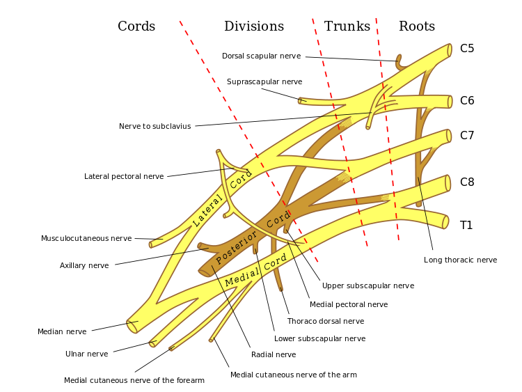
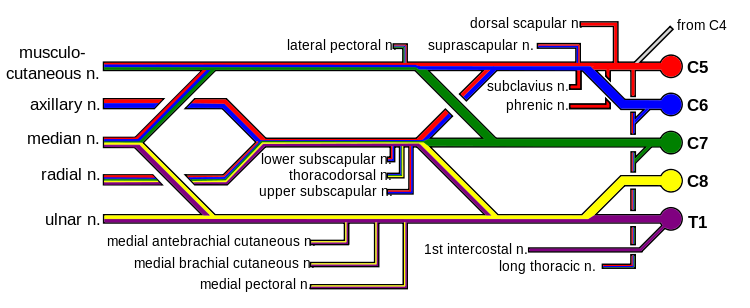
Nerves of the shoulder |
|
|
|
|
|
Dorsal
scapular |
C5 |
Off C5 root
(ventral ramus) |
Rhomboids |
|
|
Long
thoracic |
C5-C7 |
Ventral rami of
roots |
Serratus anterior |
|
|
Suprascapular
nerve |
C5, C6 |
Only branch of the
upper trunk of brachial plexus |
Supraspinatus Infraspinatus |
AC joint Glenohumeral
joint |
|
Subscapular
– upper |
|
|
|
|
|
Subscapular
- lower |
|
|
|
|
|
|
|
|
|
|
|
Nerves of the upper limb |
|
|
|
|
|
Nerve |
Roots |
Plexus |
Motor |
Sensory |
|
|
|
|
|
|
|
Axillary |
C5-6 |
Superior trunk Posterior
division Posterior cord |
Deltoid |
Patch on lateral
arm |
|
Musculocutaneous |
C5-6 |
Superior trunk Lateral cord |
Biceps Brachialis |
Lateral forearm
(Lateral cutaneous nerve of the forearm/Lateral antebrachial) |
|
Median |
C5-T1 |
Mid/Lower trunk Anterior
divisions Lateral/Med Cords |
Pronator teres Flexor carpi
radialis Palmaris longus Flexor digitorum
superficialis Anterior
interosseous nerve -
Flexor
digitorum profundus -
Flexor
pollicis longus -
Pronator
quadratus Hand -
Lumbricals -
Opponens pollicis -
Abductor
pollicis brevis -
Flexor
pollicis brevis |
Lateral 3.5
digits Palm (palmar
branch) |
|
Ulnar |
C8-T1 |
Lower trunk Anterior division Medial Cord |
Flexor carpi ulnaris Flexor digitorum profundus Hand -
Interossei -
Lumbricals
(medial) -
Abductor digiti minimi -
Oppones digiti minimi -
Flexor digiti minimi -
Adductor
pollicis -
+/- Flexor
pollicis brevis |
Median 1.5 digits Palm (palmar
branch) Dorsum of hand (dorsal
branch) |
|
Radial |
C5-C8 |
Mid/Lower trunk Posterior
Divisions Posterior Cords |
Triceps Brachioradialis Supinator Abductor pollicis
longus Extensor -
carpi
radialis (longus and brevis) -
carpi ulnaris -
carpi
digitorum -
digit minimi -
pollicis (longus
and brevis) -
indicis |
Dorsum of hand
(lateral/snuff box) |
|
|
|
|
|
|
|
|
|
|
|
|
|
|
|
|
|
|
|
|
|
|
|
|